
Glimpses of Gut Microbes
The idea that directly observing gut microbes can give us insights into their behavior is an important part of the META center’s approach to microbiome research.This week, graduate student Matthew Jemielita with colleagues in the labs of two META PIs, Karen Guillemin and Raghuveer Parthasarathy, published a paper, Spatial and Temporal Features of the Growth of a Bacterial Species Colonizing the Zebrafish Gut, describing the growth behaviors of a very simple host-microbe system: a single bacterial species colonizing the larval zebrafish gut. Using light sheet fluorescence microscopy to obtain high-resolution, three-dimensional images over many-hour time spans, the authors were able to robustly measure for the first time microbial growth rates inside the gut of a live vertebrate. More surprisingly, they uncovered intriguing correlations between the morphology of the microbial communities and their growth behaviors, finding that densely clustered microbes grew at a much faster rate than did free individuals. The mathematical form of this growth reveals that the cluster growth is not occurring at the cluster surfaces (like the growth of a sugar crystal, or snowflake), but rather is a manifestation of increased cell division throughout the cluster’s bulk. These experiments show that bacterial shape and form can be strongly linked to growth dynamics, and point the way to parameterizations that can characterize more complex multi-species colonization – the subject of experiments presently underway.
http://vimeo.com/114808879
*Video:* Colonization of the zebrafish intestine by EGFP-labeled Aeromonas veronii. Time is
measured from the start of inoculation. Scale bar: 100 microns. From: http://mbio.asm.org/content/5/6/e01751-14.abstract
Matthew Jemielita, Michael J. Taormina, Adam R. Burns, Jennifer S. Hampton, Annah S. Rolig, Karen Guillemin, and Raghuveer Parthasarathy, “Spatial and Temporal Features of the Growth of a Bacterial Species Colonizing the Zebrafish Gut.†mBio. 5, e01751–14 (2014).
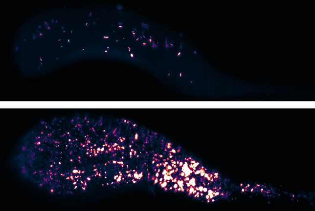
Figure Caption: Bacteria (false colored) in the gut of a live larval zebrafish. Each is a projection of a 3D image, part of a series capturing the first 16 hours of colonization of an initially empty intestine. Over this period a few intrepid individual bacteria divide into several thousand. These two images are separated in time by five hours.
December 17, 2014